Neuromuscular Pathology
 We offer comprehensive diagnostic services and timely consultations in all areas of neuromuscular pathology. For primary diagnostic workup, the muscle specimens should be submitted fresh on wet ice or snap-frozen on dry ice, while the nerve specimens should be submitted fresh on wet ice or fixed in glutaraldehyde at room temperature (full instructions can be accessed by clicking on the “Instructions and Requisition Forms” tab below). In addition, we provide second-opinion reviews and consultations for cases that were initially processed and interpreted elsewhere; the instructions for submitting previously frozen material for additional workup at UCSF are also included in the Instructions for Submission of Muscle Biopsy Consultation Specimens document.
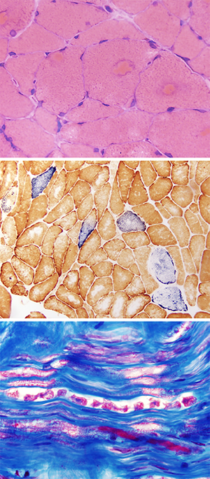
Our interpretations are based on a comprehensive panel of histochemical, immunohistochemical, and other stains (the full list is provided below); to minimize the costs, the workup starts with a limited panel of standard stains that is then supplemented by additional stains that are ordered a-la-carte as needed, based on the initial histologic findings and clinical differential diagnosis. If additional genetic and/or biochemical tests are required for a definitive diagnosis, we provide counseling on the options that are available as well as assistance with the specimen shipment to the outside reference laboratory of choice.
Standard muscle panel:
- Formalin-fixed, paraffin-embedded tissue (FFPE): H&E
- Frozen section (FS): H&E, modified Gomori Trichrome, slow/fast myosin heavy chain dual immunoperoxidase stain, NADH-TR, SDH, COX/SDH dual stain, MHC-I immunoperoxidase stain, embryonic/fetal myosin heavy chain dual immunoperoxidase stain
Standard nerve panel:
- FFPE: H&E, Congo Red
- Epon-embedded thick section: Toluidine Blue
Additional stains that can be performed a-la-carte on either specimen:
- FFPE special stains: PAS, PASD, Congo Red (and all other special stains performed by the UCSF Clinical Histology Lab)
- FFPE IPOX: dual myosin, desmin, LC3, p62/SQSTM1, TDP-43, SDHB (and all other immunoperoxidase and in situ hybridization stains performed by the UCSF Clinical Histology Lab, including stains for various microorganisms)
- FFPE immunofluorescence: kappa, lambda, IgG, IgM, IgA
- FS special stains: PAS, PASD, Sudan Black, Congo Red
- FS enzyme histochemistry: acid phosphatase, alkaline phosphatase, esterase, myophosphorylase, phosphofructokinase
- FS immunoperoxidase: CD20, CD3, CD8, CD68, C5b-9 (membrane attack complex), MHC-II, MxA (also known as Mx1), CD31, desmin, dystrophin (N terminus, C terminus, and rod domain), LC3, p62/SQSTM1, pTDP-43, single slow / fast / embryonic / fetal myosin heavy chain stains
- FS immunofluorescence: IgA, IgG, IgM, kappa, lambda
- Electron microscopy