Primary Tissue Diagnosis
Ocular Pathology (ophthalmic pathology; eye pathology) examines tissues from eye, ocular adnexa, orbit and eyelids for tumorous or non-tumorous diseases. UCSF Ocular pathology accepts tissue specimens for primary diagnosis submitted by Ophthalmologists or other pathologists. Please refer to the specimen handling for specific tissue types and diagnostic considerations. Please see shipping instructions for tissue and slides/blocks.
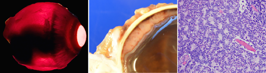
Given the complex anatomy of the eye, orbit and periorbital tissues, macroscopic evaluation of ocular pathology specimens carries utmost importance. At UCSF, macroscopic evaluations of all tissue specimens from enucleations and exenteration are performed by an ocular pathologist using specialized tools and inverted microscopes. Macroscopic examination of the tissue specimens from ocular biopsies are performed by experienced certified pathology assistants.
Histologic preparation of ocular pathology specimens also requires technical expertise and specialized tools since the specimens are either very scant or they require megablocks. UCSF Histology Laboratory provides excellent technical service combined and is equipped with state of the art tissue processors, histochemical and immunohistochemical staining technology to prepare high-quality sections and stains. We also have access to ultrastructural (electron microscopic) analysis, cytopathology services for ocular fine needle aspirates, flow cytometry and hematopathology services, in addition to a wide array of molecular diagnostics including targeted next generation sequencing [link to UCSF500].
A final pathology report is issued in Pathology reporting system, which is faxed to the referring physician and/or referring pathologist. The pathology report can also be accessed through the UCSF electronic medical record system, and through CareEverywhere